

Truth Under the Microscope: Morphological Identification of Primary Cells versus Established Cell Lines
How visual-based assessment assists researchers to judge cellular identity and avoid experimental bias
HUSTON, TX, UNITED STATES, September 10, 2026 /EINPresswire.com/ -- In daily cell-culture laboratories, the inverted phase-contrast microscope acts as investigators’ primary observational tool. Without complex staining or slide-preparation workflows, researchers can directly visualise live adherent cells through culture-vessel bottoms. Distinct morphological patterns emerge under observation: some cultures display highly diversified, intricate cellular architectures, while others show uniform, neatly arranged monolayers. These striking visual differences are far more than cosmetic features. They reveal a fundamental divide within in-vitro systems: whether cultured materials are physiologically representative primary cells, or adapted, long-term passaged cell lines. Reliable morphological evaluation serves as the first screening checkpoint to prevent misleading experimental outputs for life-science projects worldwide.
Primary cells are freshly isolated directly from native animal or human tissues including cerebral cortex and liver, and cultured for the first time. Within their limited replicative lifespan, primary cells largely retain the biological fingerprints inherited from donor tissues. Such intrinsic biological signatures are prominently reflected in cellular morphology.
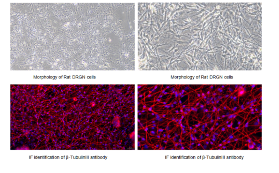
Neurons represent a classic morphological example. At culture day 3, cone-shaped or oval cell somas sprout extremely fine cellular processes. One process rapidly elongates with limited branching to become the prospective axon; multiple remaining processes repeatedly branch out as nascent dendrites. By day 7, these cellular projections interconnect and build dense web-like neural networks with electrophysiological activity.
Astrocytes exhibit distinct morphological traits: broad, irregular polygonal or star-shaped cell bodies extending abundant slender processes, comparable to the tentacles of sea stars. They frequently co-exist with neurons and function as supporting glial cells. At high density, astrocytes fuse and form continuous carpet-like monolayers.
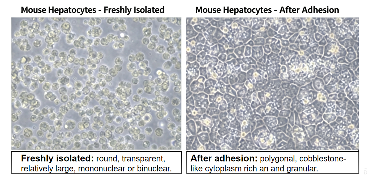
Primary hepatocytes show typical polygonal epithelial-like morphology and arrange in compact cobblestone-pattern monolayers. Their cytoplasm contains abundant lipid droplets, glycogen granules and lipofuscin deposits. Binucleated or even multinucleated phenotypes are frequently observed, a characteristic mark resulting from long-term metabolic and detoxification workload. Critically, early-passage primary hepatocytes maintain robust CYP450 enzymatic activity, a feature almost completely lost in hepatocellular carcinoma cell lines.
Figure 1 Primary Rat Dorsal Root Ganglion Neuron Cells (DRGN)
Macrophages are immune sentinels with highly plastic morphology. Under resting conditions they appear round or amoeboid with weak adherence. Once activated by stimuli such as LPS, macrophages spread rapidly and extend broad lamellipodia. They display characteristic kidney-shaped or horseshoe-shaped nuclei with ruffled, actively motile cell margins, constantly sensing surrounding microenvironments.
These diverse morphologies are not random. Primary cells retain epigenetic memory derived from in-vivo niches, preserving tissue-specific ion channels, receptor proteins and functional enzymes. Every microscopic observation reads like a biological identity card inherited from original tissues.
Cell lines: morphologically-simplified populations adapted for in-vitro culture
After the first sub-cultivation step, primary cells enter the cell-line stage. When further transformed into continuous immortalised cell lines, long-term in-vitro adaptation and genetic drift drive silent collective phenotypic remodelling, pushing cell populations toward high morphological uniformity and simplification.
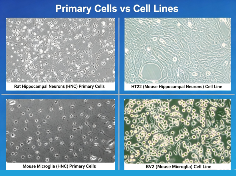
New researchers are often surprised by immortalised HT22 mouse hippocampal neuronal cells: no visible axons or dendrites can be detected under microscopy. Cells exhibit uniform spindle-shaped or near-round morphology and align in consistent orientations at high confluence. Undifferentiated SH-SY5Y human neuroblastoma cells also appear small-round or short-spindle and tend to form clusters. Such morphological convergence is widely observed across established cell lines. HeLa cells display polygonal shapes yet lose native epithelial polarity; HEK293 cells grow as uniform small-round or short-spindle populations; HepG2 hepatoma cells rarely present binucleation or cytoplasmic lipid inclusions typical of primary hepatocytes.
Morphological homogenisation signals deeper biological alterations beneath visual appearances. For instance, normal human somatic cells carry 46 chromosomes, whereas HeLa cells contain approximately 80. Elevated nucleus-to-cytoplasm ratios, intensely-stained nuclei and frequent multinucleation indicate genomic instability and accumulated mutations after extensive passaging. Many cell lines lose tissue-specific antigens and functional receptors, and shift metabolic preference from oxidative phosphorylation toward glycolysis. Proliferation becomes dominant while differentiation capacity fades. These cells become specialised for continuous culture-ware propagation yet abandon their original physiological functions.
Figure 2 Mouse primary hepatocytes. Round upon isolation, classic cobblestone morphology after attachment.
Why morphological discrepancies carry critical experimental significance
Morphological differences act as biological warning indicators, alerting researchers to one major experimental risk: loss of biological relevance.
The irreplaceable merit of primary cells lies in their close resemblance to in-vivo conditions. For blood-brain-barrier modelling, only primary brain microvascular endothelial cells can generate sufficiently high trans-endothelial electrical resistance and intact tight-junction structures. Immortalised cell lines show minimal expression of key transporters such as P-glycoprotein. In hepatic toxicology assessment, intact CYP450 enzyme systems preserved within primary hepatocytes are essential for reliable drug-metabolism evaluation.
Cell lines possess their own obvious strengths: fast proliferation and low operational cost make them suitable for high-throughput screening and large-scale gene-editing experiments. Nevertheless, phenotypic drift induced by repeated passaging may generate divergent results across different passage numbers. Morphological observation constitutes the earliest alert system for potential experimental bias.
Figure 3 Primary Cells vs. Cells Lines
Reading morphology to understand cellular behaviour
Experienced cell-culture practitioners adopt a practical morphological diagnostic workflow:
-Overall population pattern: Primary-cell cultures frequently form spreading monolayers or vortex-like arrangements; cell-line cultures appear highly uniform, dense and direction-aligned.
-Cell boundary features: Primary-cell borders are irregular with filopodia; cell-line edges tend to be smooth with consistent cellular contours.
-Nucleus-to-cytoplasm ratio and nuclear shape: Enlarged, dark-stained or multinucleated phenotypes strongly suggest cell-line populations; moderate nucleus-cytoplasm proportions with distinct nucleoli favour primary-cell identity.
-Intracellular granules and vacuoles: Primary cells commonly contain lysosomes and lipofuscin; cell-line cytoplasm looks comparatively “clean”.
-Growth characteristics: Primary cells proliferate slowly with heterogeneous populations; cell-lines multiply rapidly and reach full confluence within 2-3 days.
Morphological screening provides the first-line evaluation but is not fully conclusive. Certain primary-cell populations simplify their morphology at early passages, while some cell-line models can regain partial differentiated phenotypes after induced differentiation. Therefore, morphological assessment must be combined with specific biomarker staining — including β-Tubulin III for neurons, GFAP for astrocytes, Albumin for hepatocytes and F4/80 for macrophages — for final confirmation. Morphology delivers first-pass impressions, while biomarker staining provides formal cellular authentication. Joint interpretation enables accurate cellular identification.
Cellular morphology mirrors intrinsic cellular states, reflecting tissue origin, identity, health status and functional fate. Adjusting microscope focus and light settings represents not merely technical operations, but direct observation of biological characteristics. Morphology delivers intuitive visual evidence showing what cells are, what functions they can perform, and whether experiments rest upon biologically sound foundations.
Reading cellular morphology helps investigators interpret cell properties, which further supports correct understanding behind experimental datasets. Microscopic morphological evaluation combined with biomarker validation helps global researchers distinguish primary cells from adapted cell-line populations, minimise unexpected experimental deviation and enhance the robustness of cell-based research outputs.
About Cloud-Clone Corp.
Cloud-Clone Corp. is dedicated to the development and production of high-quality immunoassay reagents and detection solutions. With a focus on antibody engineering, multiplex assay development, and cross-platform compatibility, the company provides research tools designed to support precision medicine and advanced biomedical investigation globally. Our core products and services include the research and development of proteins, antibodies, ELISA kits, primary cells, and multiplex cytokine assay kits, as well as professional CRO services to fully meet the diverse needs of biomedical research and related fields.
For more information about Cloud-Clone Corp, visit www.cloud-clone.com.
CLOUD-CLONE CORP.(CCC)
Tel: 001-832-538-0970, 0086-27-8425-9552
Email: mail@cloud-clone.com, sales@cloud-clone.us
Visit us on social media:
LinkedIn: https://www.linkedin.com/company/cloud-clone-corp/
Instagram: https://www.instagram.com/cloudclonecorp2023/
TickTok: https://www.tiktok.com/@cloudclonecorp2026
SuKi Duan
CLOUD-CLONE CORP.WUHAN
email us here
Visit us on social media:
LinkedIn
Instagram
TikTok
X
Legal Disclaimer:
EIN Presswire provides this news content "as is" without warranty of any kind. We do not accept any responsibility or liability for the accuracy, content, images, videos, licenses, completeness, legality, or reliability of the information contained in this article. If you have any complaints or copyright issues related to this article, kindly contact the author above.
